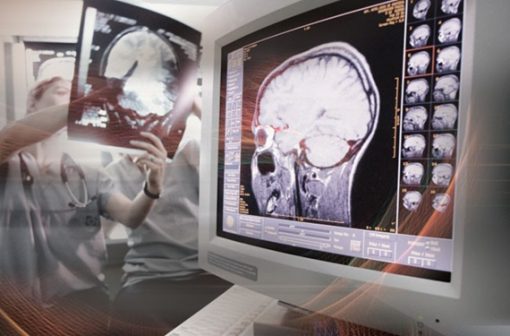
Penn Radiology’s renowned faculty provides a thorough review of best practices in Neuroradiology. Comprehensive Neuroradiology: Imaging Ideals will benefit imagers, neurologists and neurosurgeons with an extremely detailed analysis of anatomy, techniques, modalities and procedures.
Content :
Session 1: Brain I
| Brain Pain: After Hours Misses and Interpretive Errors | John H. Woo, MD |
| Brain in the Frying Pan: Imaging of Illicit Drug Abuse | Laurent LeTourneau-Guillon, MD, MSc |
| Evaluating Intra-Axial Brain Neoplasia | John H. Woo, MD |
| Hydrocephalus and CSF Flow Imaging | John H. Woo, MD |
| Early Detection: Brain Infections | John H. Woo, MD |
Session 2: Head and Neck I
| The Patient Experience in Head and Neck: Patient Facing Radiology | Laurie A. Loevner, MD |
| Cervical Lymph Node Review | Jillian W. Lazor, MD |
| Orbital Imaging Cases | Laurie A. Loevner, MD |
| The Pineal Region | Jeffrey B. Ware, MD |
| The Patient Experience in Head and Neck: Patient Facing Radiology | Laurie A. Loevner, MD |
Session 3: Spine I
| Spine Trauma Without the Drama | Jae W. Song, MD, MS |
| Spine Refined: After Hours Misses and Interpretive Errors | Jae W. Song, MD, MS |
| Degenerative Spine Disease | John H. Woo, MD |
| Imaging the Post-Operative Spine | John H. Woo, MD |
| Questions and Answers |
Session 4: Stroke
| Imaging of Spontaneous Intracerebral Hemorrhage | Laurent LeTourneau-Guillon, MD, MSc |
| Stroke Mimics: The Great Masquerade | Laurent LeTourneau-Guillon, MD, MSc |
| Patterns of Acute Arterial Stroke | Jae W. Song, MD, MS |
| Imaging of Acute Stroke: An Evolving Landscape | Piya Saraiya, MD |
| Questions and Answers |
Session 5: Brain II
| Systemic Diseases and Their Effects on the Brain | Ilya M. Nasrallah, MD, PhD |
| Nontraumatic Neurologic Emergencies | Ronald L. Wolf, MD, PhD |
| Neurodegenerative Diseases | Ilya M. Nasrallah, MD, PhD |
| Infections of the CNS | Suyash Mohan, MD |
| Discovering Adult Brain Tumors | Jeffrey B. Ware, MD |
| Advanced Surgical Planning: DTI Tractography and fMRI | Ronald L. Wolf, MD, PhD |
Session 6: Head and Neck II
| Thyroid Nodules and When to Do CT/MRI in Thyroid Cancer | Laurie A. Loevner, MD |
| EBV Associated Neoplasia of the Head and Neck | Laurie A. Loevner, MD |
| Neck is a Wreck: Case Based Review | Laurie A. Loevner, MD |
| Blind Spots in the Neck: Misses and Interpretive Errors | Laurie A. Loevner, MD |
Session 7: Spine II
| Imaging of Cervical Spine Trauma | J. Eric Schmitt, MD, PhD |
| Non-Traumatic Spine Emergencies | Robert M. Kurtz, MD |
| Spinal Neoplasia | Jae W. Song, MD, MS |
| Bone Radiology for the Brain Radiologist | Robert M. Kurtz, MD |
Session 8: Pediatrics
| Pediatric Brain Tumors | Erin Simon Schwartz, MD, FACR |
| Neuroimaging of Abusive Trauma | Karuna Shekdar, MD |
| Pediatric Spine Tumors | Karuna Shekdar, MD |
| Phakomatoses | Arastoo Vossough Modarress, MD, PhD |
Session 9: Vascular
| Cerebrovascular Anatomy (Arterial and Venous) | Robert Hurst, MD |
| Intracranial Venous Thrombosis | Jae W. Song, MD, MS |
| Vascular Malformations of the Brain | Seyed Ali Nabavizadeh, MD |
| Cerebral Aneurysms: Concepts and Controversies | Linda Bagley, MD |
| Intracranial Vessel Wall Imaging | Jae W. Song, MD, MS |
| Normal Variants – Don’t Touch Lesions | Seyed Ali Nabavizadeh, MD |
| Questions and Answers |
Session 10: Head and Neck III
| The Larynx: Patterns of Tumor Spread | Laurie A. Loevner, MD |
| PET/CT in the Head and Neck: Pearls and Pitfalls | Laurie A. Loevner, MD |
| The Anterior Skull Base Sinonasal Neoplasia With an Emphasis on Resectability | Laurie A. Loevner, MD |
| Brachial Plexus: An Easy Approach to the Nexus | Alvand Hassankhani, MD |
| One Bite at a Time: TMJ MR Imaging | Laurie A. Loevner, MD |
| Questions and Answers |
Release date: November 20, 2022
Expiration date: November 19, 2025

