UCSF Body Imaging – Abdominal and Thoracic
Learn From a World Leading Institution
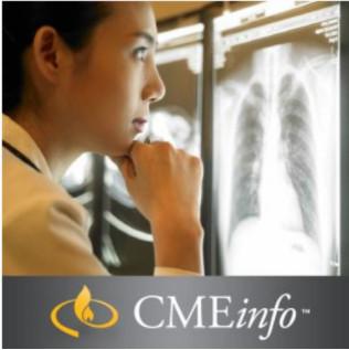
Designed for practicing radiologists working in all areas of private practice or academia, this program features recorded lectures from distinguished professors and practicing radiologists with the University of California San Francisco School of Medicine. Upon completion of this course, you will know how to:
-
-
- Differentiate common liver and pancreatic masses and pulmonary nodules
- Recognize typical and atypical appearances of lung cancer including infiltrative lung diseases
- Evaluate the small and large bowel in patients with abdominal pain
-
Wide-Ranging Clinical Update
Lectures included in this review are designed to improve interpretive and diagnostic skills related to common and uncommon conditions and diseases throughout the chest and abdomen. Available online, you can learn at your own pace in your home or office and earn up to 18.25 AMA PRA Category 1 Credits ™. It’s an ideal way to stay current and incorporate new guidelines into your daily practice.
PET and Oncology
-
-
- A Comprehensive Approach to Lung Cancer Cases – David M. Naeger, MD
- Complete PET/CT Assessment of Pulmonary Nodules – David M. Naeger, MD
- Workshop: Incidentials and Artifacts on PET/CT: How to Not Overcall – David M. Naeger, MD
-
- Imaging Workup of Cystic Pancreatic Masses – Judy Yee, MD, FACR
- Problem Solving Solid Pancreatic Masses – Judy Yee, MD, FACR
-
Abdominal Imaging
-
-
- Uterus and Post-Menopausal Bleeding – Ruth B. Goldstein, MD
-
- CT Colonography: Pearls, Pitfalls, Current Practice – Judy Yee, MD, FACR
- Workshop: Abdominal Imaging: Challenging Cases – Judy Yee, MD, FACR
-
- Focal Liver Masses – Benjamin M. Yeh, MD
- Keys for Acute Bowel Imaging – Benjamin M. Yeh, MD
-
Efficient CT and US in Practice
-
-
- Can We Biopsy Fewer Nodules? – Ruth B. Goldstein, MD
- Complications of Early Pregnancy, Focus on New Guidelines – Ruth B. Goldstein, MD
- Fetal CNS: What Are You Detecting, What Are You Missing? – Ruth B. Goldstein, MD
-
- Vascular Pearls for Improved Abdominopelvic Interpretation – Benjamin M. Yeh, MD
- Workshop: Maximizing Your CT Scans Value – Benjamin M. Yeh, MD
-
Thoracic Imaging
-
- Acute Aortic Syndrome – Brett M. Elicker, MD
- Emergent Cardiac Findings on Routine Chest CT – Brett M. Elicker, MD
- Update on Pulmonary Embolism – Brett M. Elicker, MD
- Workshop: Challenging Chest Cases from the Reading Room – Brett M. Elicker, MD
-
- The Myriad Appearances of Lung Cancer: Everything You Need to Know – David M. Naeger, MD
-
- Airway Disease – Part 1: Trachea and Large Airways – W. Richard Webb, MD
- Airway Disease – Part 2: Small Airways – W. Richard Webb, MD
- Infiltrative Lung Diseases – Part 1: Case-Based Review – W. Richard Webb, MD
- Infiltrative Lung Diseases – Part 2: Case-Based Review – W. Richard Webb, MD
Accreditation
The University of California, San Francisco School of Medicine (UCSF) is accredited by the Accreditation Council for Continuing Medical Education to provide continuing medical education for physicians.
Designation
UCSF designates this enduring material for a maximum of 18.25 AMA PRA Category 1 Credits ™. Physicians should claim only the credit commensurate with the extent of their participation in the activity.
The total credits are inclusive of 13.0 in CT, 1.5 in MR, 3.0 in OB/GYN US and 0.75 in PET/CT.
Series Release: May 16, 2015
Series Expiration: May 15, 2018 (deadline to register for credit)CME credit is obtained upon successful completion of a multiple-choice self-test and evaluation (included with the series), as well as an additional $45 processing fee. CME Credit registration forms must be submitted prior to series expiration date, certificates will be dated upon receipt and cannot be dated retroactively.
Learning Objectives
Upon completion of this course, you will know how to:
- Differentiate common solitary liver masses
- Recognize typical and atypical appearances of lung cancer and pancreatic masses
- Describe the proper management of a renal incidentaloma
- Evaluate the small and large bowel in patients with abdominal pain
- Utilize ultrasound to assess fetal development
- Identify artifacts on PET/CT
- Differentiate treatment effects from tumor recurrence after liver-directed anti-tumor therapy
- Recommend appropriate uses of CT angiography
-
Intended Audience
UCSF Body Imaging: Abdominal & Thoracic is intended for radiologists in both clinical practice and academic environments, and addresses important topics in gastrointestinal, pulmonary, thoracic, oncologic and ob-gyn imaging.
Only logged in customers who have purchased this product may leave a review.
Related Products
Internal Medicine Videos
VIDEO MEDICAL
VIDEO MEDICAL
VIDEO MEDICAL
VIDEO MEDICAL
VIDEO MEDICAL
VIDEO MEDICAL
VIDEO MEDICAL
VIDEO MEDICAL
VIDEO MEDICAL
VIDEO MEDICAL
VIDEO MEDICAL
VIDEO MEDICAL
VIDEO MEDICAL
VIDEO MEDICAL
VIDEO MEDICAL
VIDEO MEDICAL
VIDEO MEDICAL
VIDEO MEDICAL
VIDEO MEDICAL
VIDEO MEDICAL
VIDEO MEDICAL
VIDEO MEDICAL
VIDEO MEDICAL
VIDEO MEDICAL
VIDEO MEDICAL
VIDEO MEDICAL
VIDEO MEDICAL
Classic Lectures in Pathology: What You Need to Know: Neuropathology – A Video CME Teaching Activity
VIDEO MEDICAL
VIDEO MEDICAL
VIDEO MEDICAL
VIDEO MEDICAL
VIDEO MEDICAL
VIDEO MEDICAL
VIDEO MEDICAL
VIDEO MEDICAL
VIDEO MEDICAL
VIDEO MEDICAL
VIDEO MEDICAL
VIDEO MEDICAL
VIDEO MEDICAL
VIDEO MEDICAL
VIDEO MEDICAL
VIDEO MEDICAL
VIDEO MEDICAL
VIDEO MEDICAL
VIDEO MEDICAL
VIDEO MEDICAL
VIDEO MEDICAL

























Reviews
There are no reviews yet.