- NEW Illustration overviews highlight the autonomic nerves to clarify nerve and muscle innervations.
- NEW and updated images reflect the latest clinical insights through:
- 15 NEW Illustrations
- 160 revised figures
- NEW and updated surface anatomy images
- NEW pulses photos
- Renowned, high-resolution, dynamically colored illustrations organized in dissection sequence enable the formation of 3-D constructs for each body region and provide detailed, realistic reference during dissection.
- Tables detail muscles, vessels, and other anatomic information in an easy-to-use format ideal for review and study.
- Enhanced medical imaging includes more than 100 clinically significant MRIs, CT images, ultrasound scans, and corresponding orientation drawings to help students confidently apply the laboratory experience to clinical rotations.
- Color schematic illustrations reinforce the relationships of structures and anatomical concepts in vibrant detail.
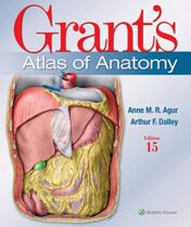
Grant’s Atlas of Anatomy
For more than seventy-five years, Grant’s Atlas of Anatomy has maintained a tradition of excellence while continually adapting to meet the needs of each generation of students. The updated fifteenth edition is a visually stunning reference that delivers the accuracy, pedagogy, and clinical relevance expected of this classic atlas, with new and enhanced features that make it even more practical and user-friendly. Illustrations drawn from real specimens, presented in surface-to-deep dissection sequence, set Grant’s Atlas of Anatomy apart as the most accurate reference available for learning human anatomy. These realistic representations bring structures to life and provide students with the ultimate lab resource.
PAGES: 896
YEAR: 2020
PUBLICATION: LWW
LANGUAGE: English
Related Products
Anatomy Books
$25
$12
$40
Anatomy Books
$13
$12
Anatomy Books
$12
Anatomy Books
$15
Anatomy Books
$15
Anatomy Books
$15
Anatomy Books
$15
$15
Anatomy Books
$15
Anatomy Books
$14
$12
$15
$17
Anatomy Books
$15
$14
Anatomy Books
$10
$20
Anatomy Books
$22
$14
$20
Anatomy Books
$12
Anatomy Books
$14
Anatomy Books
$15
Anatomy Books
$15
$15
$15
Anatomy Books
$15
Anatomy Books
$15
Anatomy Books
$15
Anatomy Books
$15
$15
Anatomy Books
$12
$9
$12

