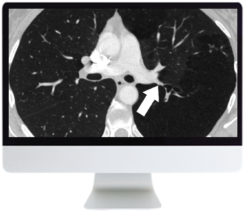
ARRS Basic Chest Imaging 2019 (CME VIDEOS)
This course covers basic topics in thoracic imaging, a rapidly evolving subspecialty. Course content provides a focused review of basic chest imaging topics, as well as important updates regarding thoracic disease entities and technological advancements. Topics include imaging of airways, imaging of atelectasis, imaging of infections in immunocompromised hosts, current concepts and approach to management of pulmonary nodules, as well as imaging of pulmonary embolism.
Learning Outcomes and Lectures
After completing this course, the learner should be able to:
- Recognize anatomic appearances of atelectasis, differentiate among etiologies, and describe imaging signs
- Discuss imaging appearance and differential diagnosis of infectious and noninfectious causes of airspace disease in immunosuppressed patients
- Discuss key factors in nodule management, management approach focusing on current guidelines, and pitfalls
- Review the spectrum of adenocarcinoma, correlation of CT with pathology, and impact on subsolid nodule management
- Discuss patterns of multiple small nodules, anatomic basis for pattern-based approach, strategies for diagnosis, common causes, and more rare considerations
- Recognize challenges related to pulmonary embolism diagnosis with CT pulmonary angiogram, identify solutions, and evaluate the problem of overdiagnosisReview the imaging of large and small airways, protocols, tracheal and bronchial tumors, and tracheomalacia and bronchiectasis.
Module 1
- Spectrum of Atelectasis on Chest Radiographs and CT—J. Czum
- Imaging of Infections in Immunocompromised Hosts—R. Madan
- Management of Solid Pulmonary Nodules: Characterization and Update—J. Ko
- Adenocarcinoma and the Subsolid Nodule: Current Concepts—J. Ko
Module 2
- Nodular Pattern on Chest CT—D. Manos
- Pulmonary Embolism: Beyond Filling Defects—Technique, Pitfalls, Misdiagnosis, and Overdiagnosis—L. Haramati
- Imaging of Airways—M. Hammer
Related Products
Radiology Books
American journal of Neuroradiology 2023 Full Archives (True PDF)
Radiology Books
Radiology Books
Radiology Books
American Journal of Roentogelogy 2023 Full Archives (True PDF)
Radiology Books
Contemporary Diagnostic Radiology 2023 Full Archives (True PDF)
Radiology Books
Radiology Books
Advances in Medical Imaging, Detection, and Diagnosis (EPUB)
ORTHOPAEDICS SURGERY
PLASTIC & RECONSTRUCTIVE SURGERY
The Aesthetic Society Nuances in Injectables The Next Beauty Frontier 2022
Radiology Books
Radiology Books
Radiology Books
Tumor Imaging: CT Colonography Online Course 2022 (CME VIDEOS)
Radiology Books
Radiology Books
Radiology Books
Radiology Books
Multimodality Imaging, Volume 1 (Original PDF from Publisher)
Radiology Books
Absolute Breast Imaging Review (Original PDF from Publisher)
Radiology Books
Radiology Books
American journal of Neuroradiology 2022 Full Archives (True PDF)
Radiology Books
How to Read a Normal Scan : Brain CT (High Quality Image PDF)
Radiology Books
How to read a Normal Scan: Spine MRI (High Quality Image PDF)
Radiology Books
How to Read a Normal Scan : SPINE CT (High Quality Image PDF)
Radiology Books
Radiology Books
Fuzzy Sets Methods in Image Processing and Understanding (EPUB)
Radiology Books
Radiology Books
Radiology Books
Radiology Books
Radiology Books
Imaging for Students, 5th Edition (Original PDF from Publisher)
Radiology Books
Radiology Books
Eco-Doppler Vascular: Guia Prático (Original PDF from Publisher)
Radiology Books
Contemporary Diagnostic Radiology 2021 Full Archives (True PDF)
Radiology Books
American Journal of Roentogelogy 2022 Full Archives (True PDF)
Radiology Books
Radiology Books
Radiology Books
Radiology Books
Radiology Books
Top 3 Differentials in Pediatric Radiology: A Case Review (EPUB)
Radiology Books
Top 3 Differentials in Nuclear Medicine: A Case Review (EPUB)

