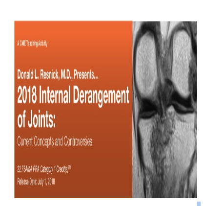
Donald L. Resnick, M.D. Presents: 2018 Internal Derangement of Joints: Current Concepts and Controversies (CME VIDEOS)
$60
31 Video Files (.mp4 format).+1PDF
File Size = 4.83 GB
| TARGET AUDIENCE This CME activity provides an update of current information on MR Imaging in the assessment of musculoskeletal disorders. Essential anatomy, physiology and pathology are emphasized that explain imaging findings in disorders of the shoulder, elbow, wrist, hand, hip, knee, ankle and foot. MR imaging findings in the assessment of common problems in peripheral joints are compared to those derived from other imaging methods. |
Physicians: Educational Symposia is accredited by the Accreditation Council for Continuing Medical Education (ACCME) to provide continuing medical education for physicians.Educational Symposia designates this enduring material for a maximum of 22.75 AMA PRA Category 1 Credit(s)TM. Physicians should claim only the credit commensurate with the extent of their participation in the activity.Credits awarded for these enduring activities are designated “SA-CME” by the American Board of Radiology (ABR) and qualify toward fulfilling requirements for Maintenance of Certification (MOC) Part II: Lifelong Learning and Self-assessment.
All activity participants are required to take a written or online test in order to be awarded credit. (Exam materials, if ordered, will be sent with your order.) All course participants will also have the opportunity to critically evaluate the program as it relates to practice relevance and educational objectives.
AMA PRA Category 1 Credit(s)TM for these programs may be claimed until June 30, 2021.
This CME activity was planned and produced by Educational Symposia, the leader in diagnostic imaging education since 1975.
This CME activity was planned and produced in accordance with the ACCME Essential Areas and Elements.
TOPICS/SPEAKERS
– Common and Uncommon Disorders of Synovium-Lined Joints
– Osteochondral, Subchondral, Cortical: Acute Injuries
Tears and Pseudotears, Impingement, and Calcification
-Brachialis, Triceps, and Flexor and Extensor Tendons
Related Products
ORTHOPAEDICS SURGERY
ORTHOPAEDICS SURGERY
ORTHOPAEDICS SURGERY
ORTHOPAEDICS SURGERY
Master Techniques in Orthopaedic Surgery: The Foot and Ankle, 4th Edition (Videos)
ORTHOPAEDICS SURGERY
Master Techniques in Orthopaedic Surgery: The Foot and Ankle, 4th Edition EPUB
ORTHOPAEDICS SURGERY
ORTHOPAEDICS SURGERY
Synopsis of Foot and Ankle Surgery (Original PDF from Publisher)
ORTHOPAEDICS SURGERY
Orthopaedic Knowledge Update®: Musculoskeletal Tumors 5 (EPUB)
ORTHOPAEDICS SURGERY
Primer on the Metabolic Bone Diseases and Disorders of Mineral Metabolism, 10th edition (EPUB)
ORTHOPAEDICS SURGERY
TMJ Extremity Proficiency Product 2023 – ExtremityExperts (Videos)
ORTHOPAEDICS SURGERY
ORTHOPAEDICS SURGERY
Frozen Shoulder: Present and Future (Original PDF from Publisher)
ORTHOPAEDICS SURGERY
Quand et comment reprendre une prothèse de genou ? (French Edition) (True PDF from Publisher)
ORTHOPAEDICS SURGERY
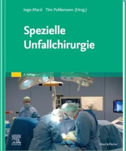
Spezielle Unfallchirurgie (German Edition), 2nd Edition (True PDF from Publisher)
ORTHOPAEDICS SURGERY
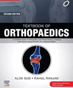
Textbook of Orthopaedics, 2nd Edition (True PDF from Publisher)
ORTHOPAEDICS SURGERY
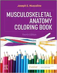
Musculoskeletal Anatomy Coloring Book, 4th Edition (True PDF from Publisher)
ORTHOPAEDICS SURGERY
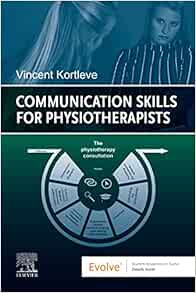
Communication Skills for Physiotherapists (True PDF from Publisher)
ORTHOPAEDICS SURGERY
Trauma Surgery Essentials: A Must-Know Guide to Emergency Management (True PDF from Publisher)
ORTHOPAEDICS SURGERY
Complications in Orthopaedics: Pediatrics (True PDF from Publisher)
ORTHOPAEDICS SURGERY
Dupuytren’s Disease: A Scientific Review (True PDF from Publisher)
ORTHOPAEDICS SURGERY
Knee Pain in Sports Medicine: Essentials of Diagnosis and Treatment (True PDF from Publisher)
ORTHOPAEDICS SURGERY
Campbell’s Core Orthopaedic Procedures, 2nd Edition (True PDF from Publisher)
ORTHOPAEDICS SURGERY
Surgical Anatomy and Exposures of the Knee: A Surgical Atlas (Original PDF from Publisher)
ORTHOPAEDICS SURGERY
ORTHOPAEDICS SURGERY
Lower Extremity Proficiency Product 2023 – ExtremityExperts (Videos)
ORTHOPAEDICS SURGERY
CTS Extremity Proficiency Product 2023 – ExtremityExperts (Videos)
ORTHOPAEDICS SURGERY
Orthopaedic Physical Therapy Secrets, 4th Edition (True PDF from Publisher)
ORTHOPAEDICS SURGERY
Laboratory Manual for Clinical Kinesiology and Anatomy, 5th Edition (EPUB)
ORTHOPAEDICS SURGERY
Complications in Orthopaedics: Spine Surgery (EPUB + Converted PDF)
ORTHOPAEDICS SURGERY
ORTHOPAEDICS SURGERY
Plastic Surgery: Hand and Upper Limb, Volume 5, 5th Edition (True PDF from Publisher)
ORTHOPAEDICS SURGERY
Netter. Sistema musculoesquelético integrado (True PDF from Publisher)
ORTHOPAEDICS SURGERY
Evidence-Based Physical Therapy for the Pelvic Floor, 3rd Edition (True PDF from Publisher)
ORTHOPAEDICS SURGERY
Neuromuscular Case Studies, 2nd Edition (True PDF from Publisher)
ORTHOPAEDICS SURGERY
Pflege konkret Chirurgie Orthopädie Urologie, 6th Edition (True PDF from Publisher)
ORTHOPAEDICS SURGERY
Emergency and Trauma Care for Nurses and Paramedics, 4th Edition (True PDF from Publisher)
ORTHOPAEDICS SURGERY
ORTHOPAEDICS SURGERY
Thieme Dissector Volume 2: Abdomen and Lower Limb, 2nd Edition (Original PDF from Publisher)
ORTHOPAEDICS SURGERY
Thieme Dissector Volume 1: Upper Limb and Thorax, 2nd Edition (Original PDF from Publisher)
ORTHOPAEDICS SURGERY
Imagerie musculosquelettique traumatique (Original PDF from Publisher)
ORTHOPAEDICS SURGERY
Screening in der Physiotherapie, 2nd Edition (Original PDF from Publisher)
ORTHOPAEDICS SURGERY
ORTHOPAEDICS SURGERY
ORTHOPAEDICS SURGERY
Stoller’s Orthopaedics and Sports Medicine: The Hip (Original PDF from Publisher)
ORTHOPAEDICS SURGERY
Rockwood and Green’s Fractures in Adults, 10th edition (EPUB + Converted PDF)
ORTHOPAEDICS SURGERY
Moderne Wundversorgung, 10th Edition (German Edition) (True PDF from Publisher)
ORTHOPAEDICS SURGERY
Pflegewissen Wunden, 3rd Edition (German Edition) (True PDF from Publisher)
ORTHOPAEDICS SURGERY
ORTHOPAEDICS SURGERY
Revision Total Knee Arthroplasty, 3rd edition (Original PDF from Publisher)
ORTHOPAEDICS SURGERY
ORTHOPAEDICS SURGERY
Soft Tissue Therapy for the Lower Limb (Original PDF from Publisher)
ORTHOPAEDICS SURGERY
ORTHOPAEDICS SURGERY
Atención inicial al trauma grave: Las primeras 24 horas (Original PDF from Publisher)
ORTHOPAEDICS SURGERY
Spinal Deformities: The Comprehensive Text, 2nd edition (Original PDF from Publisher+Videos)
ORTHOPAEDICS SURGERY
Handbook of Spine Surgery, 3rd Edition + Videos (Original PDF from Publisher)
ORTHOPAEDICS SURGERY
Rockwood and Wilkins’ Fractures in Children, 10th edition (EPUB + Converted PDF)
ORTHOPAEDICS SURGERY
ORTHOPAEDICS SURGERY
Intracranial and Spinal Dural Arteriovenous Fistulas (Original PDF from Publisher)
ORTHOPAEDICS SURGERY
Balanced Ligamentous Tension in Osteopathic Practice (Original PDF from Publisher)

