Atlas of Pathologic Myopia 2020 Original pdf+videos
$15
Atlas of Pathologic Myopia 2020 Original pdf+videos
This Atlas provides many beautiful images obtained with state-of-the-art technologies, including optical coherence tomography (OCT), OCT angiography, fundus autofluorescence, and wide-field fundus imaging, as well as traditional images and fluorescein/ICG angiograms. Gathered at the world’s largest High Myopia Clinic, the images are based on the long-term follow-up data of more than 6,000 patients from Japan and abroad.
Related Products
Ophthalmology & Optometry Books
Ophthalmology & Optometry Books
Atlas of Clinical and Surgical Orbital Anatomy, 3rd edition (ePub+Converted PDF)
Ophthalmology & Optometry Books
Ophthalmology & Optometry Books
Ophthalmology & Optometry Books
Optical Coherence Tomography Angiography of the Eye (Original PDF from Publisher)
Ophthalmology & Optometry Books
Everyday OCT: A Handbook for Clinicians and Technicians, 2nd Edition (EPUB)
Ophthalmology & Optometry Books
Ophthalmology & Optometry Books
Anti-VEGF Use in Ophthalmology (Original PDF from Publisher)
Ophthalmology & Optometry Books
Subjective Refraction and Prescribing Glasses, 3rd Edition (Original PDF from Publisher)
Ophthalmology & Optometry Books
Parsons’ Diseases of the Eye, 24th edition (Original PDF from Publisher)
Ophthalmology & Optometry Books
Ophthalmology & Optometry Books
Kline’s Neuro-Ophthalmology Review Manual, 8th Edition (EPUB)
Ophthalmology & Optometry Books
Simple: The Inner Game of Ophthalmic Practice Success, 2nd Edition (Original PDF from Publisher)
Ophthalmology & Optometry Books
Ophthalmology & Optometry Books
Ophthalmology & Optometry Books
Ophthalmology & Optometry Books
Optical Coherence Tomography of Ocular Diseases, 4th Edition (EPUB)
Ophthalmology & Optometry Books
Ophthalmology & Optometry Books
Curbside Consultation in Cornea and External Disease: 49 Clinical Questions, 2nd Edition (EPUB)
Ophthalmology & Optometry Books
Essential Business Fundamentals for the Successful Eye Care Practice (Original PDF from Publisher)
Ophthalmology & Optometry Books
Dry Eye Disease: A Practical Guide (Original PDF from Publisher)
Ophthalmology & Optometry Books
Ophthalmology & Optometry Books
Ophthalmology & Optometry Books
Ophthalmology & Optometry Books
Ophthalmology & Optometry Books
Optical Coherence Tomography Angiography Atlas (Original PDF from Publisher)
Ophthalmology & Optometry Books
Ophthalmology & Optometry Books
Ophthalmology & Optometry Books
Ophthalmology & Optometry Books
Ophthalmology & Optometry Books
Ophthalmology & Optometry Books
Ophthalmology & Optometry Books
Certified Ophthalmic Technician Exam Review Manual, 3rd Edition (EPUB)
Ophthalmology & Optometry Books
American Journal of Ophthalmology 2023 Full Archives (True PDF)
Ophthalmology & Optometry Books
British Journal of Ophthalmology 2023 Full Archives (True PDF)
Ophthalmology & Optometry Books
Simple: The Inner Game of Ophthalmic Practice Success, 2nd Edition (EPUB)
Ophthalmology & Optometry Books
Therapeutic Targets for Diabetic Retinopathy: A Translational Approach (Original PDF from Publisher)
Ophthalmology & Optometry Books
Ophthalmology & Optometry Books
EMQs in Ophthalmology, Dermatology and ENT (Original PDF from Publisher)
Ophthalmology & Optometry Books
Photo Acoustic and Optical Coherence Tomography Imaging, Volume 2 (Original PDF from Publisher)
Ophthalmology & Optometry Books
Retinal Detachment Surgery and Proliferative Vitreoretinopathy, 2nd Edition (EPUB)
Ophthalmology & Optometry Books
Ophthalmology & Optometry Books
Optical Coherence Tomography of the Anterior Segment (Original PDF from Publisher)
Ophthalmology & Optometry Books
Global Perspectives in Ocular Oncology (Original PDF from Publisher)
Ophthalmology & Optometry Books
Neuro-Ophthalmology: Case Based Practice (Original PDF from Publisher)
Ophthalmology & Optometry Books
Ophthalmology & Optometry Books
Ophthalmology & Optometry Books
Ophthalmology & Optometry Books
Ophthalmology & Optometry Books
Ophthalmology & Optometry Books
Ophthalmology & Optometry Books
Ophthalmology & Optometry Books
Ophthalmology & Optometry Books
Ophthalmology & Optometry Books
Ophthalmology & Optometry Books
Ophthalmology & Optometry Books
Ophthalmology & Optometry Books
Low Vision: Principles and Management (Original PDF from Publisher)
Ophthalmology & Optometry Books
Ophthalmology & Optometry Books
Atlas do Fundo de Olho, 2nd Edition (Original PDF from Publisher)
Ophthalmology & Optometry Books
Secrets: Oftalmologia em Cores – Perguntas e Respostas, 4th Edition (Original PDF from Publisher)
Ophthalmology & Optometry Books
Interdisciplinary Management of Orbital Diseases: Textbook and Atlas,1st edition (EPUB)
Ophthalmology & Optometry Books
HEAD AND NECK SURGERY & OTOLARYNGOLOGY
Manual of Eye, Ear, Nose, and Throat Emergencies (Original PDF from Publisher)
Ophthalmology & Optometry Books
Ophthalmology & Optometry Books
Ophthalmology & Optometry Books
Ophthalmology & Optometry Books
Ophthalmology & Optometry Books
Ophthalmology Review: A Case-Study Approach, 2nd Edition (EPUB)
Ophthalmology & Optometry Books
Ophthalmology & Optometry Books
Ophthalmology & Optometry Books
Clinical Pathways in Neuro-Ophthalmology: An Evidence-Based Approach (EPUB)
Ophthalmology & Optometry Books
Siga a Seta – Oculoplástica em Fluxogramas: Guia Prático de Oftalmologia da USP (EPUB)
Ophthalmology & Optometry Books
The Art of Pterygium Surgery: Mastering Techniques and Optimizing Results (EPUB)
Ophthalmology & Optometry Books
Strabismus Surgery: Innovative and Classic Approaches (EPUB)
Ophthalmology & Optometry Books
Pediatric Ophthalmology Surgery and Procedures: Tricks of the Trade (EPUB)
Ophthalmology & Optometry Books
Ophthalmology & Optometry Books
The Art of Refractive Cataract Surgery: For Residents, Fellows, and Beginners (EPUB)
Ophthalmology & Optometry Books
Pre-Descemet’s Endothelial Keratoplasty (PDEK) (Original PDF from Publisher+Videos)
Ophthalmology & Optometry Books
Current Opinion in Ophthalmology 2022 Full Archives (True PDF)
Ophthalmology & Optometry Books
Kanskis Klinische Ophthalmologie: Ein systematischer Ansatz, 9th Edition (EPUB3)
Ophthalmology & Optometry Books
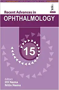
Recent Advances in Opthalmology 15 (Original PDF from Publisher)
Ophthalmology & Optometry Books
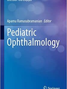
Pediatric Ophthalmology (Current Practices in Ophthalmology) (Original PDF from Publisher)
Ophthalmology & Optometry Books
Pediatric Ophthalmology (Current Practices in Ophthalmology) (EPUB)
Ophthalmology & Optometry Books
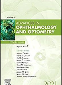
Advances in Ophthalmology and Optometry 2021 (Original PDF from Publisher)
Ophthalmology & Optometry Books
Advances in Ophthalmology and Optometry 2022 (Original PDF from Publisher)
Ophthalmology & Optometry Books
Advances in Ophthalmology and Optometry 2018 (Original PDF from Publisher)
Ophthalmology & Optometry Books
Advances in Ophthalmology and Optometry 2017 (Original PDF from Publisher)
Ophthalmology & Optometry Books
Dynamic Light Scattering Spectroscopy of the Human Eye (Original PDF from Publisher)
Ophthalmology & Optometry Books
Dynamic Light Scattering Spectroscopy of the Human Eye (EPUB)
Ophthalmology & Optometry Books
Ophthalmology & Optometry Books
Ophthalmology & Optometry Books
Gems of Ophthalmology—Cataract Surgery (Original PDF from Publisher)
Ophthalmology & Optometry Books
Introduction to Visual Optics: A Light Approach (Original PDF from Publisher)
Ophthalmology & Optometry Books
Ophthalmology & Optometry Books
Atlas of Oculoplastic and Orbital Surgery, 2nd edition (Original PDF from Publisher)
Ophthalmology & Optometry Books
Common Eye Diseases and their Management, 5th Edition (Original PDF from Publisher)
Ophthalmology & Optometry Books
Common Eye Diseases and their Management, 5th Edition (EPUB)
Ophthalmology & Optometry Books
Clinical Pediatric Ophthalmology and Strabismus (Original PDF from Publisher)

