- Language : English
- ISBN-10 : 939055313X
- ISBN-13 : 978-9390553136
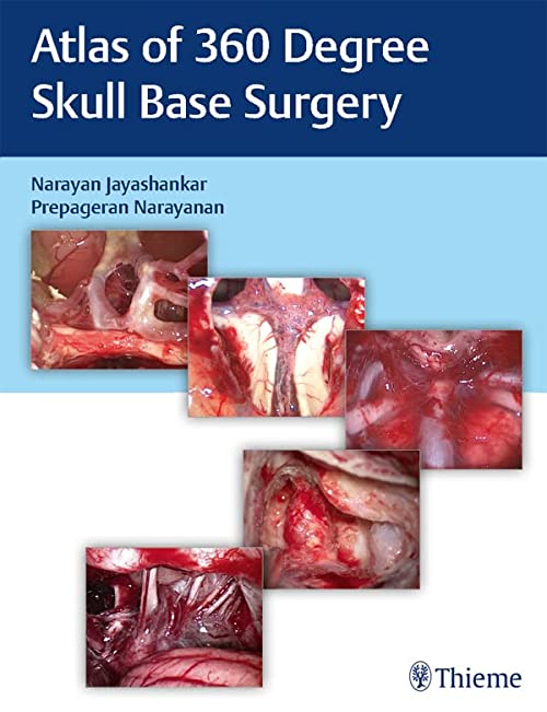
Atlas of 360 degrees Skull Base Surgery PDF
The sellar region contains numerous essential structures, which embryologically arise in a complex manner, resulting in tight neighborship of otherwise unrelated tissues. As the prime example, the pituitary gland contains epithelial structures, arising from the future oral cavity, differentiates into neuroendocrine cells, has a dedicated vascular network facilitating hormone release and at the same time comprises a direct junction to the central nervous system (CNS). Besides the pituitary gland, the sellar region also lies within the former anterior neuropore where germ cell progenitors are located in the embryo and at the same time is close to the anterior end of the former notochord, a cartilage-like structure still present in the early human embryo and evolution’s first attempt toward bilateral symmetric creatures with a spinal cord that would later evolve into vertebrates and ultimately humans. Such a potpourri of highly specialized and very different progenitor cell lineages explains the often-confusing morphological appearance of tumors that most likely arise from these structures that typically have a midline orientation. Besides lesions caused by nonresident cell populations or external factors, such as meningiomas, metastases, apoplexy, or abscesses, the sellar region is highly enriched in rare, locoregionally specific neoplasms of extremely diverse cell-of-origin lineage.
Section I: Pituitary Gland Tumors
Chapter 1 Pathologies of the Sellar Region
Introduction to the Sellar Region
Introduction to the Pituitary Gland
Pathologies and Its Variants
Anatomical Considerations
Clinical Considerations
Radiological Considerations
Surgical Considerations
Preoperative Considerations
Selection of the Surgical Approach
Presurgical Phase
Nasal Phase (Tailored Approach)
Osteodural Phase (Zonal Dissection)
Intradural Phase (Tumor Resection)
Skull Base Defect Closure
Postoperative Care
Nonsurgical Treatment Modalities
Management of Complications
Tips and Pearls
Acknowledgments
References
Chapter 2 Endoscopic Transnasal Approach for Pituitary Tumors
Chapter 3 Endoscopic Endonasal Surgeries for Pituitary Macroadenoma
Chapter 4 Endoscopic Endonasal Revision Surgery for Pituitary Adenoma
Chapter 5 Endoscopic Endonasal Surgery for Functioning Pituitary Adenoma
Chapter 6 Endoscopic Endonasal Surgery for Cystic Lesions of the Sella
Section II: Supra-sellar Region
Section III: Transcribriform Approach
Section IV: Clival Region
Section V: Craniocervical Junction
Section VI: Orbital Region
Section VII: Orbital Apex and Optic Nerve
Section VIII: Infratemporal Fossa and Parapharyngeal Space
Section IX: Petrous Region
Section X: Meckel’s Cave
Section XI: Cerebrospinal Fluid Rhinorrhea
Section XII: Reconstruction in Endoscopic Skull Base
Section XIII: Cavernous Sinus
Section XIV: Sinonasal Tumors
Section XV: Skullbase Fibro-osseous Pathologies
Section XVI: Complications
Section XVII: Basics in Otology
Section XVIII: Translabyrinthine Approach
Section XIX: Approaches Through the Otic Capsule
Section XX: Retrolabyrinthine Approach
Section XXI: Presigmoid Approach
Section XXII: Management of Glomus Tumors/Approaches to Jugular Foramen
Section XXIII: Approach to Foramen Magnum Tumors
Section XXIV: Surgeries for Vertigo
Section XXV: Microvascular Decompression
Section XXVI: Retrosigmoid Approach
Section XXVII: Middle Cranial Fossa
Section XXVIII: Otogenic Infections and Tumors
Section XXIX: Rehabilitative Surgery
Atlas of 360 Degree Skull Base Surgery
Product details
Related Products
Neurosurgery books PDF
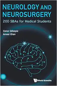
Neurology and Neurosurgery: 200 Sbas for Medical Students (Original PDF from Publisher)
Neurosurgery books PDF
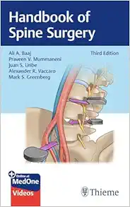
Handbook of Spine Surgery, 3rd Edition + Videos (Original PDF from Publisher)
Neurosurgery books PDF
Spinal Deformities: The Comprehensive Text, 2nd edition (Original PDF from Publisher+Videos)
Neurosurgery books PDF
Journal of Neurosurgical Anesthesiology 2024 Full Archives (True PDF)
Neurosurgery books PDF
Neurosurgery books PDF
Neurosurgery books PDF
Operative Techniques: Spine Surgery, 4th edition (Original PDF from Publisher)
Neurosurgery books PDF
Neurosurgery books PDF
Anterior Controllable Antedisplacement and Fusion: Technique in Spinal Surgery (EPUB)
Neurosurgery books PDF
Neurosurgery books PDF
Neurosurgery books PDF
Neurosurgery books PDF
Neurosurgery books PDF
Biomechanics of Spine Stabilization: Self-Assessment and Review (Original PDF from Publisher)
Neurosurgery books PDF
Neurosurgery books PDF
Neurosurgery books PDF
Lyftogt Perineural Injection Treatment: How to Treat Peripheral Nerve Pain
Neurosurgery books PDF
Neurosurgery books PDF
Spinal Deformities: The Comprehensive Text 2nd Edition PDF & VIDEO
Neurosurgery books PDF
Neurosurgery books PDF
Neurosurgery books PDF
Journal Of Neurosurgery Pediatrics 2023 Full Archives (True PDF)
Neurosurgery books PDF
Journal Of NeuroInterventional Surgery 2023 Full Archives (True PDF)
Neurosurgery books PDF
External Ventricular And Lumbar Drains: Indications, Procedures, And Patient Care (EPUB)
Neurosurgery books PDF
Journal Of Neurosurgical Anesthesiology 2023 Full Archives (True PDF)
Neurosurgery books PDF
Neuroimaging Clinics Of North America 2023 Full Archives (True PDF)
Neurosurgery books PDF
Neuroimaging Clinics Of North America 2022 Full Archives (True PDF)
Neurosurgery books PDF
Neurosurgery Clinics Of North America 2023 Full Archives (True PDF)
Neurosurgery books PDF
Neurosurgery Clinics Of North America 2022 Full Archives (True PDF)
Neurosurgery books PDF
An Inner Approach To Cranial Osteopathy (Original PDF From Publisher)
Neurosurgery books PDF
Neurosurgery books PDF
Neurosurgery books PDF
Neurosurgery books PDF
Neurosurgery books PDF
Neurosurgery books PDF
Neurosurgery books PDF
Neurosurgery books PDF
Manual De Neurocirugia (2 Volumenes, 9ª Edicion) (High Quality Image PDF)
Neurosurgery books PDF
Neurosurgery books PDF
Neurosurgery books PDF
Neurosurgery books PDF
Intracranial Arteriovenous Malformations: Essentials for Patients and Practitioners
Neurosurgery books PDF
Neuro-Oncology Compendium for the Boards and Clinical Practice
Neurosurgery books PDF
Skull Base Reconstruction: Management of Cerebrospinal Fluid Leaks and Skull Base Defects
Neurosurgery books PDF
Neurosurgery books PDF
Neurosurgery books PDF
Neurosurgery books PDF
Master Techniques in Orthopaedic Surgery: The Spine, 4th Edition
Neurosurgery books PDF
Ultrasonography of the Prenatal Brain, Third Edition Original PDF
Neurosurgery books PDF
Neurosurgery books PDF
Neurosurgery books PDF
Neurosurgery books PDF
Neurosurgery books PDF
Neurosurgery books PDF
Neurosurgery books PDF
Surgery of the Cerebellopontine Angle, 2nd Edition (Original PDF from Publisher)
Neurosurgery books PDF
Surgical Nuances of Head Injury (Original PDF from Publisher)
Neurosurgery books PDF
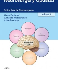
Neurosurgery Updates Critical Care for Neurosurgeons Volume 3 (Original PDF from Publisher)
Neurosurgery books PDF
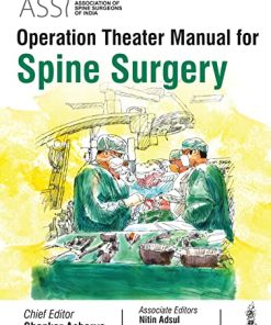
ASSI Operation Theater Manual for Spine Surgery (Original PDF from Publisher)
Neurosurgery books PDF
Journal of Neurosurgery: Spine 2022 Full Archives (True PDF)
Neurosurgery books PDF
Journal of Neurosurgery: Pediatrics 2022 Full Archives (True PDF)
Neurosurgery books PDF
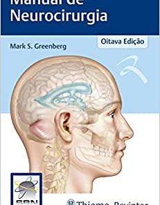
Manual de Neurocirurgia, 8th Edition (Original PDF from Publisher)
Neurosurgery books PDF
The Comprehensive Neurosurgery Board Preparation Book: Illustrated Questions and Answers (EPUB)
Neurosurgery books PDF
Neurosurgical Operative Atlas: Spine and Peripheral Nerves, 3rd Edition (EPUB)
HEAD AND NECK SURGERY & OTOLARYNGOLOGY
Atlas of Facial Nerve Surgeries and Reanimation Procedures Original PDF
Neurosurgery books PDF
Neurosurgery books PDF
Carotid Treatment: Principles and Techniques, 3rd Edition 2023 Original PDF
Neurosurgery books PDF
Condutas em Neurocirurgia: Fundamentos Práticos – Crânio (EPUB)
Neurosurgery books PDF
Neurosurgery books PDF
Neurosurgery books PDF
Transnasal Endoscopic Skull Base and Brain Surgery: Surgical Anatomy and its Applications (EPUB)
Neurosurgery books PDF
Surgery of the Thoracic Spine: Principles and Techniques (EPUB)
Neurosurgery books PDF
Meningiomas of the Skull Base: Treatment Nuances in Contemporary Neurosurgery (EPUB)
Neurosurgery books PDF
Cirurgia Endoscópica da Coluna Vertebral, 2nd Edition (Original PDF from Publisher)

