- Format: 120 Video Files (.mp4 format).
- File Size: 5.03 GB.
-
Note : We will send ebook download link after confirmation of payment via paypal success
Payment methods: Visa or master card (Paypal)
123sonography – Vascular Lower Extremity BachelorClass 2019
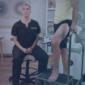
With the Vascular Lower Extremity BachelorClass you will gain a strong foundation and comprehensive introduction to performing and interpreting vascular ultrasound examinations.
The Vascular Lower Extremity BachelorClass is designed to provide you with a comprehensive introduction to lower extremity vascular sonography by guiding you through the essentials of establishing scan protocols as well as the lower extremity anatomy and its vascular ultrasound characteristics.
You will be trained to translate 3D anatomy to 2D images with emphasis placed on a thorough understanding of the principles underlying the Doppler examination and clinical applications using Color Doppler and Spectral Doppler techniques. Learn to identify normal venous and arterial anatomy during peripheral lower extremity ultrasound imaging while demonstrating the confidence to incorporate protocols, techniques & interpretation criteria to improve diagnostic and treatment accuracy in your practice. You will also learn about the relations between acoustic principles, haemodynamics, and the sonographic representation of major vessels and blood flow.
The Vascular Lower Extremity BachelorClass covers multiple indications that may be present in your patients’ examination, such as cardiac output, dehydration and infection, which can all affect the vascular system in different ways and aggravate existing conditions.
All of this knowledge will be made practical and easy to follow with several case studies to ensure a strong foundational understanding of a wide range of vascular conditions.
Topics/Speaker:
01.01 Course Introduction
01.02 Ultrasound System- Basic Overview
01.03 Ultrasound System- Comprehensive Overview
01.04 Transducer Selection
01.05 Setting Up the Ultrasound Environment
01.06 Image Orientation
01.07 Transducer Ergonomics
01.08 Transducer Manipulation
01.09 Basic Knobology
01.10 Image Optimization- B-Mode
01.11 Image Optimization- Color Doppler
01.12 Image Optimization- Spectral Doppler
02.01 Sonographer-Physician Communication
02.02 Sonographer-Patient Communication
03.01 Lower Extremity Arterial Anatomy
03.02 Lower Extremity Arterial Physiology
03.03 Lower Extremity Arterial Hemodynamics
04.01 Atherosclerosis
04.02 Aneurysm
04.03 Fibromuscular Dysplasia
04.04 Congenital Anomalies
05.01 Patient Positioning and Transducer Selection
05.02 Proximal Common Femoral Artery
05.03 Distal Common Femoral Artery
05.04 Superficial Femoral Artery
05.05 Popliteal Artery
05.06 Proximal Tibioperoneal Trunk
05.07 Distal Tibioperoneal Trunk
05.08 Calf Anatomy
05.09 Posterior Tibial Artery
05.10 Peroneal Artery
05.11 Anterior Tibial Artery
05.12 Dorsalis Pedis Artery
05.13 Calf Arteries
06.01 Clinical Session-Superficial Femoral Artery
06.02 Clinical Session-Above Knee Level
06.03 Clinical Session-Below Knee Level
06.04 Case Study
06.05 Clinical Session-Common Femoral to Deep Femoral Arteries
06.06 Clinical Session-Superficial Femoral Artery
06.07 Case Study
06.08 Patient Story
06.09 Clinical Session-Superficial Femoral Artery
06.10 Clinical Session-Above Knee Level
06.11 Clinical Session-Aneurysm Assessment
06.12 Case Study
06.13 Case Study
06.14 Case Study-Aortoiliac Occlusion
07.01 Lower Extremity Venous Anatomy
07.02 Lower Extremity Venous Physiology
07.03 Lower Extremity Venous Hemodynamics
08.01 Venous Thromboembolism and Deep Vein Thrombosis
08.02 Varicose Veins
08.03 Venous Severity Measurement
09.01 Patient Positioning and Transducer Selection
09.02 Common Femoral Vein
09.03 Saphenofemoral Junction
09.04 Femoral Vein
09.05 Popliteal Vein
09.06 Intramuscular Veins
09.07 Posterior Tibial and Peroneal Veins
09.08 Anterior Tibial Veins
09.09 Great Saphenous Vein
09.10 Small Saphenous Vein
10.01 Examination Overview
10.02 Patient Positioning and Transducer Selection
10.03 Common Femoral Vein
10.04 Femoral Vein
10.05 Popliteal Vein
10.06 Calf Veins
10.07 Great Saphenous Veins
10.08 Small Saphenous Veins
10.09 Patient Positioning
10.10 Common Femoral Vein
10.11 Femoral Vein
10.12 Popliteal Vein
10.13 Great Saphenous Vein- Saphenofemoral Junction
10.14 Great Saphenous Vein- Terminal Valve
10.15 Great Saphenous Vein- Subterminal Valve
10.16 Great Saphenous Vein- Proximal Thigh Level
10.17 Great Saphenous Vein- Distal Thigh Level
10.18 Great Saphenous Vein- Knee Level
10.19 Great Saphenous Vein- Below Knee Level
10.20 Great Saphenous Vein- Proximal Calf Level
10.21 Great Saphenous Vein- Distal Calf Level
10.22 Tributaries
10.23 Anterior Accessory Saphenous Vein
10.24 Posterior Accessory Saphenous Vein
10.25 Intersaphenous Connection
10.26 Small Saphenous Vein- Below Knee Level
10.27 Small Saphenous Vein- Proximal – Mid Calf Level
10.28 Small Saphenous Vein- Distal Calf Level
10.29 Conclusion
11.01 Clinical Session- Venous Patency Evaluation
11.02 Clinical Session- Common Femoral Vein
11.03 Clinical Session- Great Saphenous Vein
11.04 Clinical Session- Tributary and Perforator
11.05 Case Study
11.06 Patient Story
11.07 Clinical Session- Contralateral Common Femoral Vein
11.08 Clinical Session- Common Femoral Vein
11.09 Clinical Session- Femoral Vein
11.10 Clinical Session- Popliteal Vein
11.11 Clinical Session- Calf Veins
11.12 Three Case Studies- Saphenous Vein Insufficiency
11.13 Case Study- The Heart and the Vascular System
11.14 Case Study- Deep Vein Thrombosis
11.15 Case Study- Massive Deep Vein Thrombosis
12.01 Ankle-Brachial Index- Examination Overview
12.02 Ankle-Brachial Index- Cuff Placement
12.03 Ankle-Brachial Index- Brachial Pressures
12.04 Ankle-Brachial Index- Ankle Pressures
12.05 Ankle-Brachial Index- Review of Findings
12.06 Segmental Examination- Segmental Pressures
12.07 Segmental Examination- Review of Findings
12.08 Pulse-Volume Recording
12.09 Continuous Wave Doppler
12.10 Toe-Brachial Index
12.11 Plethysmography Testing
12.12 Intravascular Ultrasound
Product Details
Related Products
CARDIOLOGY BOOKS
CARDIOLOGY BOOKS
Internal Medicine Videos
2022 AANEM Spring Virtual Conference Collection 2022 (CME VIDEOS)
Internal Medicine Videos
The International Congress Of Parkinson and Movement Disorder 2022 (MDS Congress) (CME VIDEOS)
GENERAL PEDIATRICS
Chestnet Pediatric Pulmonary Board Review On Demand 2022 (CME VIDEOS)
INTENSIVE CARE BOOKS
Chestnet Critical Care Board Review On Demand 2022 (CME VIDEOS)
Internal Medicine Books
Internal Medicine Books
The PassMachine Medical Oncology Board Review 2020 (v5.1) (Beattheboards) (Lectures)
Internal Medicine Books
8th Congress of the European Academy of Neurology – Europe 2022 (CME VIDEOS)
Internal Medicine Books
MD Anderson A Comprehensive Board Review in Hematology and Medical Oncology 2021 (CME VIDEOS)
Internal Medicine Videos
CARDIOLOGY BOOKS
Mayo Clinic Echocardiography Online Board Review 2022 (CME VIDEOS)
Internal Medicine Videos
Internal Medicine Books
Internal Medicine Books
The PassMachine Addiction Medicine Board Review 2022 (v3.1) (Beattheboards) (Lectures)
Internal Medicine Videos
Internal Medicine Videos
Internal Medicine Videos
Internal Medicine Videos
The Brigham Board Review and Comprehensive Update in Rheumatology 2022 (CME VIDEOS)
Internal Medicine Videos
Contemporary Issues in Breast Pathology uscap 2022 Items Included in the Purchase of this Course
Internal Medicine Videos
Internal Medicine Videos
Internal Medicine Videos
Internal Medicine Videos
Internal Medicine Videos
CHEST Advanced Critical Care Echocardiography Board Review Exam Course Virtual Event 2020
Internal Medicine Videos
Internal Medicine Videos
Internal Medicine Videos
Internal Medicine Videos
Cleveland Clinic Digestive Disease and Surgery Update OnDemand 2019
Internal Medicine Videos
GI BOARD REVIEW (The William M. Steinberg Board Review in Gastroenterology)
Internal Medicine Videos
Internal Medicine Videos
Johns Hopkins Review of Medical and Surgical Gastroenterology 2018 (Videos+PDFs)
Internal Medicine Videos
Internal Medicine Videos
Internal Medicine Videos
USCAP Tutorial in Pathology of the GI Tract, Pancreas and Liver 2019
Internal Medicine Videos
Internal Medicine Videos
The Brigham Board Review in Gastroenterology and Hepatology 2018
Internal Medicine Videos
Internal Medicine Videos
2019 Classic Lectures in Pathology What You Need to Know Pancreatobiliary Pathology
Internal Medicine Videos
2019 Classic Lectures in Pathology What You Need to Know Gastrointestinal Pathology
Internal Medicine Videos
Internal Medicine Videos
Internal Medicine Videos
Internal Medicine Videos
Internal Medicine Videos
AAFP FAMILY MEDICINE BOARD REVIEW SELF-STUDY PACKAGE – 13TH EDITION 2020
Internal Medicine Videos
Internal Medicine Videos
Internal Medicine Videos
Internal Medicine Videos
Internal Medicine Videos
Internal Medicine Videos
Internal Medicine Videos
Internal Medicine Videos
Internal Medicine Videos
Internal Medicine Videos
2019 Association for Community Health Improvement (ACHI) National Conference
Internal Medicine Videos
A Core Curriculum in Adult Primary Care Medicine 2018-2019 Lecture Series
Internal Medicine Videos
2019 Classic Lectures in Pathology What You Need to Know Endocrine Pathology
Internal Medicine Videos
Introduction to Adult EM Bootcamp + The Practice of Emergency Medicine (Hippo) 2020
Internal Medicine Videos
CCME The National Emergency Medicine Board Review course 2020
Internal Medicine Videos
Internal Medicine Videos
Internal Medicine Videos
Internal Medicine Videos
Internal Medicine Videos
ISICEM International Symposium on Intensive Care & Emergency Medicine 2020
Internal Medicine Videos
CCME Emergency Medicine & Acute Care: A Critical Appraisal Series 2020
Internal Medicine Videos
Internal Medicine Videos
Internal Medicine Videos
Internal Medicine Videos
CCME National Emergency Medicine Board Review Self-Study 2019 (Videos)
Internal Medicine Videos
Internal Medicine Videos
Internal Medicine Videos
The Passmachine Critical Care Medicine Board Review Course 2018
Internal Medicine Videos
Internal Medicine Videos
Internal Medicine Videos
Internal Medicine Videos
Internal Medicine Videos
Internal Medicine Videos
Internal Medicine Videos
Internal Medicine Videos
Internal Medicine Videos
Internal Medicine Videos
Internal Medicine Videos
The National Family Medicine Board Review Self-Study Course 2020
Internal Medicine Videos
The Brigham and Dana-Farber Board Review in Hematology and Oncology 2020 (Videos+PDFs)
Internal Medicine Videos
Internal Medicine Videos
Internal Medicine Videos
Internal Medicine Videos
The Brigham Board Review in Pulmonary Medicine 2020 (Videos+PDFs)
Internal Medicine Videos
Internal Medicine Videos
Internal Medicine Videos
Internal Medicine Videos
Internal Medicine Videos
Internal Medicine Videos
36th Annual UCLA Intensive Course in Geriatric Medicine and Board Review 2020 (Videos+PDFs)
Internal Medicine Videos
Need-to-Know Emergency Medicine: A Review for Physicians in a Hurry 2020 (Videos+PDFs)
Internal Medicine Videos
Internal Medicine Videos

