- Relate MR image findings of the shoulder and knee to clinically relevant orthopaedic disorders
- Develop optimized protocols for imaging of the musculoskeletal system, including protocols used to suppress metal hardware in patients with joint replacements
- Understand the MRI anatomy of the shoulder, wrist, hip, knee, ankle, fingers and toes
- Identify abnormalities of the joints, muscles, bone marrow and cartilage on MRI and their clinical significance
- Recognize common bone tumors with a pertinent differential diagnosis
- Identify suitable protocols and recognize abnormalities related to cervical spine trauma
- Understand peripheral nerve imaging protocols and pertinent findings
- MRI of the Postoperative Shoulder – Lynne S. Steinbach, MD
- Pitfalls in Knee MRI – Lynne S. Steinbach, MD
- Pseudotumors – Lynne S. Steinbach, MD
- Digital MRI: the Fingers & Toes – John F. Feller, MD
- MRI Following Joint Arthroplasty – John F. Feller, MD
- Imaging of Cervical Spine Trauma – John F. Feller, MD
- Pitfalls in Shoulder MRI – Daria Motamedi, MD
- MRI of the Hip: Labrum and FAI – Daria Motamedi, MD
- MRI of the Wrist: Ligaments and TFCC – Daria Motamedi, MD
- Cartilage and Osteochondral Disease – Thomas M. Link, MD, PhD
- Knee ACL – Thomas M. Link, MD, PhD
- Ankle MRI – Thomas M. Link, MD, PhD
- Bone Tumors – Thomas M. Link, MD, PhD
- MRI of the Hip: Tendons – Christine B. Chung, MD
- Patterns of Bone Marrow Edema – Christine B. Chung, MD
- Rotator Cuff and Impingement – Christine B. Chung, MD
- Knee Meniscus – Christine B. Chung, MD
- Peripheral Nerve Imaging – Christine B. Chung, MD
UCSF Musculoskeletal MR Imaging 2016 – Videos + PDFs
Series Release: May 1, 2016
Series Expiration: April 30, 2019 (deadline to register for credit)
File Size : 3.56Gb
University of California San Francisco Clinical Update
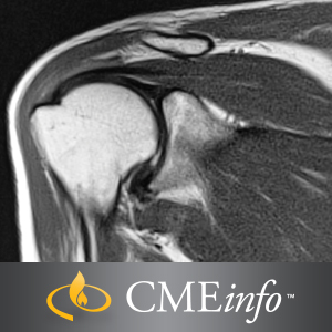
This comprehensive, state-of-the-art update focuses on different aspects of musculoskeletal MR imaging. Special attention is directed to bone marrow, muscle and tendon disorders, and nerve entrapment.
An Acclaimed Review of Musculoskeletal MR Imaging
This comprehensive, state-of-the-art update focuses on different aspects of musculoskeletal MR imaging. Along with a review of the anatomy and pathology of each joint, speakers discuss evaluation of cartilage lesions and the newest sequences, optimized protocols with 1.5T and 3T MR units, and differential diagnoses of bone and soft-tissue tumors. Special attention is directed to bone marrow, muscle and tendon disorders, and nerve entrapment. This program will help you to better:
Topics/Speakers
Note : We will send ebook download link after confirmation of payment via paypal success
Payment methods: Visa or master card (Paypal)
Related Products
ORTHOPAEDICS SURGERY
ORTHOPAEDICS SURGERY
ORTHOPAEDICS SURGERY
ORTHOPAEDICS SURGERY
Master Techniques in Orthopaedic Surgery: The Foot and Ankle, 4th Edition (Videos)
ORTHOPAEDICS SURGERY
Master Techniques in Orthopaedic Surgery: The Foot and Ankle, 4th Edition EPUB
ORTHOPAEDICS SURGERY
ORTHOPAEDICS SURGERY
Synopsis of Foot and Ankle Surgery (Original PDF from Publisher)
ORTHOPAEDICS SURGERY
Orthopaedic Knowledge Update®: Musculoskeletal Tumors 5 (EPUB)
ORTHOPAEDICS SURGERY
Primer on the Metabolic Bone Diseases and Disorders of Mineral Metabolism, 10th edition (EPUB)
ORTHOPAEDICS SURGERY
TMJ Extremity Proficiency Product 2023 – ExtremityExperts (Videos)
ORTHOPAEDICS SURGERY
ORTHOPAEDICS SURGERY
Frozen Shoulder: Present and Future (Original PDF from Publisher)
ORTHOPAEDICS SURGERY
Quand et comment reprendre une prothèse de genou ? (French Edition) (True PDF from Publisher)
ORTHOPAEDICS SURGERY
Spezielle Unfallchirurgie (German Edition), 2nd Edition (True PDF from Publisher)
ORTHOPAEDICS SURGERY
Textbook of Orthopaedics, 2nd Edition (True PDF from Publisher)
ORTHOPAEDICS SURGERY
Musculoskeletal Anatomy Coloring Book, 4th Edition (True PDF from Publisher)
ORTHOPAEDICS SURGERY
Communication Skills for Physiotherapists (True PDF from Publisher)
ORTHOPAEDICS SURGERY
Trauma Surgery Essentials: A Must-Know Guide to Emergency Management (True PDF from Publisher)
ORTHOPAEDICS SURGERY
Complications in Orthopaedics: Pediatrics (True PDF from Publisher)
ORTHOPAEDICS SURGERY
Dupuytren’s Disease: A Scientific Review (True PDF from Publisher)
ORTHOPAEDICS SURGERY
Knee Pain in Sports Medicine: Essentials of Diagnosis and Treatment (True PDF from Publisher)
ORTHOPAEDICS SURGERY
Campbell’s Core Orthopaedic Procedures, 2nd Edition (True PDF from Publisher)
ORTHOPAEDICS SURGERY
Surgical Anatomy and Exposures of the Knee: A Surgical Atlas (Original PDF from Publisher)
ORTHOPAEDICS SURGERY
ORTHOPAEDICS SURGERY
Lower Extremity Proficiency Product 2023 – ExtremityExperts (Videos)
ORTHOPAEDICS SURGERY
CTS Extremity Proficiency Product 2023 – ExtremityExperts (Videos)
ORTHOPAEDICS SURGERY
Orthopaedic Physical Therapy Secrets, 4th Edition (True PDF from Publisher)
ORTHOPAEDICS SURGERY
Laboratory Manual for Clinical Kinesiology and Anatomy, 5th Edition (EPUB)
ORTHOPAEDICS SURGERY
Complications in Orthopaedics: Spine Surgery (EPUB + Converted PDF)
ORTHOPAEDICS SURGERY
ORTHOPAEDICS SURGERY
Plastic Surgery: Hand and Upper Limb, Volume 5, 5th Edition (True PDF from Publisher)
ORTHOPAEDICS SURGERY
Netter. Sistema musculoesquelético integrado (True PDF from Publisher)
ORTHOPAEDICS SURGERY
Evidence-Based Physical Therapy for the Pelvic Floor, 3rd Edition (True PDF from Publisher)
ORTHOPAEDICS SURGERY
Neuromuscular Case Studies, 2nd Edition (True PDF from Publisher)
ORTHOPAEDICS SURGERY
Pflege konkret Chirurgie Orthopädie Urologie, 6th Edition (True PDF from Publisher)
ORTHOPAEDICS SURGERY
Emergency and Trauma Care for Nurses and Paramedics, 4th Edition (True PDF from Publisher)
ORTHOPAEDICS SURGERY
ORTHOPAEDICS SURGERY
Thieme Dissector Volume 2: Abdomen and Lower Limb, 2nd Edition (Original PDF from Publisher)
ORTHOPAEDICS SURGERY
Thieme Dissector Volume 1: Upper Limb and Thorax, 2nd Edition (Original PDF from Publisher)
ORTHOPAEDICS SURGERY
Imagerie musculosquelettique traumatique (Original PDF from Publisher)
ORTHOPAEDICS SURGERY
Screening in der Physiotherapie, 2nd Edition (Original PDF from Publisher)
ORTHOPAEDICS SURGERY
ORTHOPAEDICS SURGERY
ORTHOPAEDICS SURGERY
Stoller’s Orthopaedics and Sports Medicine: The Hip (Original PDF from Publisher)
ORTHOPAEDICS SURGERY
Rockwood and Green’s Fractures in Adults, 10th edition (EPUB + Converted PDF)
ORTHOPAEDICS SURGERY
Moderne Wundversorgung, 10th Edition (German Edition) (True PDF from Publisher)
ORTHOPAEDICS SURGERY
Pflegewissen Wunden, 3rd Edition (German Edition) (True PDF from Publisher)
ORTHOPAEDICS SURGERY
ORTHOPAEDICS SURGERY
Revision Total Knee Arthroplasty, 3rd edition (Original PDF from Publisher)
ORTHOPAEDICS SURGERY
ORTHOPAEDICS SURGERY
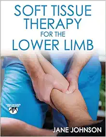
Soft Tissue Therapy for the Lower Limb (Original PDF from Publisher)
ORTHOPAEDICS SURGERY
ORTHOPAEDICS SURGERY
Atención inicial al trauma grave: Las primeras 24 horas (Original PDF from Publisher)
ORTHOPAEDICS SURGERY
Spinal Deformities: The Comprehensive Text, 2nd edition (Original PDF from Publisher+Videos)
ORTHOPAEDICS SURGERY
Handbook of Spine Surgery, 3rd Edition + Videos (Original PDF from Publisher)
ORTHOPAEDICS SURGERY
Rockwood and Wilkins’ Fractures in Children, 10th edition (EPUB + Converted PDF)
ORTHOPAEDICS SURGERY
ORTHOPAEDICS SURGERY
Intracranial and Spinal Dural Arteriovenous Fistulas (Original PDF from Publisher)
ORTHOPAEDICS SURGERY
Balanced Ligamentous Tension in Osteopathic Practice (Original PDF from Publisher)

