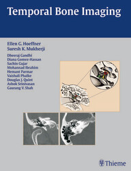
Temporal Bone Imaging 1st Edition PDF
$15
by Ellen G. Hoeffner (Editor), Suresh Kumar Mukherji (Editor)
- Each chapter features succinct descriptions of epidemiology, clinical features, pathology, treatment, and imaging findings for CT and MRI
- Bulleted lists of pearls highlight important imaging considerations
- More than 200 high-quality images demonstrate anatomy, pathologic concepts, as well as postoperative outcomes
- Publisher : Thieme; 1st edition (March 26, 2008)
- Language : English
- Hardcover : 244 pages
- ==========================+======================
-
Note : We will send ebook download link after confirmation of payment via paypal success
Temporal Bone Imaging 1st Edition PDF
Concise coverage of common temporal bone pathologies in a case-based format
Temporal Bone Imaging is a case-based review of the current techniques for imaging the various temporal bone pathologies frequently encountered in the clinical setting. Detailed discussion of anatomy provides essential background on the complex structure of the temporal bone, as well as the external auditory canal, middle ear and mastoid air cells, facial nerve, and inner ear. Chapters are divided into separate sections based on the anatomic location of the problem, with each chapter addressing a different disease entity.
Highlights:
This book will serve as a valuable reference and refresher for radiologists, neuroradiologists, otologists, and head and neck surgeons. Its concise, case-based presentation will help residents and fellows in radiology and otolaryngology-head and neck surgery prepare for board examinations.
Product details
Related Products
Neurosurgery books PDF
Neurology and Neurosurgery: 200 Sbas for Medical Students (Original PDF from Publisher)
Neurosurgery books PDF
Handbook of Spine Surgery, 3rd Edition + Videos (Original PDF from Publisher)
Neurosurgery books PDF
Spinal Deformities: The Comprehensive Text, 2nd edition (Original PDF from Publisher+Videos)
Neurosurgery books PDF
Journal of Neurosurgical Anesthesiology 2024 Full Archives (True PDF)
Neurosurgery books PDF
Neurosurgery books PDF
Neurosurgery books PDF
Operative Techniques: Spine Surgery, 4th edition (Original PDF from Publisher)
Neurosurgery books PDF
Neurosurgery books PDF
Anterior Controllable Antedisplacement and Fusion: Technique in Spinal Surgery (EPUB)
Neurosurgery books PDF
Neurosurgery books PDF
Neurosurgery books PDF
Neurosurgery books PDF
Neurosurgery books PDF
Biomechanics of Spine Stabilization: Self-Assessment and Review (Original PDF from Publisher)
Neurosurgery books PDF
Neurosurgery books PDF
Neurosurgery books PDF
Lyftogt Perineural Injection Treatment: How to Treat Peripheral Nerve Pain
Neurosurgery books PDF
Neurosurgery books PDF
Spinal Deformities: The Comprehensive Text 2nd Edition PDF & VIDEO
Neurosurgery books PDF
Neurosurgery books PDF
Neurosurgery books PDF
Journal Of Neurosurgery Pediatrics 2023 Full Archives (True PDF)
Neurosurgery books PDF
Journal Of NeuroInterventional Surgery 2023 Full Archives (True PDF)
Neurosurgery books PDF
External Ventricular And Lumbar Drains: Indications, Procedures, And Patient Care (EPUB)
Neurosurgery books PDF
Journal Of Neurosurgical Anesthesiology 2023 Full Archives (True PDF)
Neurosurgery books PDF
Neuroimaging Clinics Of North America 2023 Full Archives (True PDF)
Neurosurgery books PDF
Neuroimaging Clinics Of North America 2022 Full Archives (True PDF)
Neurosurgery books PDF
Neurosurgery Clinics Of North America 2023 Full Archives (True PDF)
Neurosurgery books PDF
Neurosurgery Clinics Of North America 2022 Full Archives (True PDF)
Neurosurgery books PDF
An Inner Approach To Cranial Osteopathy (Original PDF From Publisher)
Neurosurgery books PDF
Neurosurgery books PDF
Neurosurgery books PDF
Neurosurgery books PDF
Neurosurgery books PDF
Neurosurgery books PDF
Neurosurgery books PDF
Neurosurgery books PDF
Manual De Neurocirugia (2 Volumenes, 9ª Edicion) (High Quality Image PDF)
Neurosurgery books PDF
Neurosurgery books PDF
Neurosurgery books PDF
Neurosurgery books PDF
Intracranial Arteriovenous Malformations: Essentials for Patients and Practitioners
Neurosurgery books PDF
Neuro-Oncology Compendium for the Boards and Clinical Practice
Neurosurgery books PDF
Skull Base Reconstruction: Management of Cerebrospinal Fluid Leaks and Skull Base Defects
Neurosurgery books PDF
Neurosurgery books PDF
Neurosurgery books PDF
Neurosurgery books PDF
Master Techniques in Orthopaedic Surgery: The Spine, 4th Edition
Neurosurgery books PDF
Ultrasonography of the Prenatal Brain, Third Edition Original PDF
Neurosurgery books PDF
Neurosurgery books PDF
Neurosurgery books PDF
Neurosurgery books PDF
Neurosurgery books PDF
Neurosurgery books PDF
Neurosurgery books PDF
Surgery of the Cerebellopontine Angle, 2nd Edition (Original PDF from Publisher)
Neurosurgery books PDF
Surgical Nuances of Head Injury (Original PDF from Publisher)
Neurosurgery books PDF
Neurosurgery Updates Critical Care for Neurosurgeons Volume 3 (Original PDF from Publisher)
Neurosurgery books PDF
ASSI Operation Theater Manual for Spine Surgery (Original PDF from Publisher)
Neurosurgery books PDF
Journal of Neurosurgery: Spine 2022 Full Archives (True PDF)
Neurosurgery books PDF
Journal of Neurosurgery: Pediatrics 2022 Full Archives (True PDF)
Neurosurgery books PDF
Manual de Neurocirurgia, 8th Edition (Original PDF from Publisher)
Neurosurgery books PDF
The Comprehensive Neurosurgery Board Preparation Book: Illustrated Questions and Answers (EPUB)
Neurosurgery books PDF
Neurosurgical Operative Atlas: Spine and Peripheral Nerves, 3rd Edition (EPUB)
HEAD AND NECK SURGERY & OTOLARYNGOLOGY
Atlas of Facial Nerve Surgeries and Reanimation Procedures Original PDF
Neurosurgery books PDF
Neurosurgery books PDF
Carotid Treatment: Principles and Techniques, 3rd Edition 2023 Original PDF
Neurosurgery books PDF
Condutas em Neurocirurgia: Fundamentos Práticos – Crânio (EPUB)
Neurosurgery books PDF
Neurosurgery books PDF
Neurosurgery books PDF
Transnasal Endoscopic Skull Base and Brain Surgery: Surgical Anatomy and its Applications (EPUB)
Neurosurgery books PDF
Surgery of the Thoracic Spine: Principles and Techniques (EPUB)
Neurosurgery books PDF
Meningiomas of the Skull Base: Treatment Nuances in Contemporary Neurosurgery (EPUB)
Neurosurgery books PDF
Cirurgia Endoscópica da Coluna Vertebral, 2nd Edition (Original PDF from Publisher)

