- ==========================+======================
-
Note : We will send ebook download link after confirmation of payment via paypal success
Payment methods: Visa or master card (Paypal)
-
- Apply the latest knowledge to diagnoses and differentials for common musculoskeletal tumors
- Identify and differentiate various arthritides
- Diagnose osseous infections and mimics
- Recognize normal joint anatomy from pathology, and avoid common pitfalls
- Spot commonly-missed fractures
- Implement ultrasound as a useful tool in musculoskeletal imaging
- ACL Reconstruction – Matthew D. Bucknor, MD
- Helpful Approaches to Common MSK Tumors – Matthew D. Bucknor, MD
- MRI of Femoroacetabular Impingement – Matthew D. Bucknor, MD
- Osseous Infectious Dilemmas – Matthew D. Bucknor, MD
- Radiographic Checklist for Hip Impingement – Matthew D. Bucknor, MD
- Glenohumeral Joint Instability – Christine B. Chung, MD
- Metabolic and Endocrine Disorders – Christine B. Chung, MD
- MR Imaging Evaluation of Elbow Ligaments – Christine B. Chung, MD
- MR Imaging Evaluation of the Elbow Tendons – Christine B. Chung, MD
- Rotator Cuff: Basics and Beyond – Christine B. Chung, MD
- Ankle MRI – Thomas M. Link, MD, PhD
- Arthritis and Infection – Thomas M. Link, MD, PhD
- Cartilage and Osteochondral Disease – Thomas M. Link, MD, PhD
- Arthroplasty Complications – Kevin C. McGill, MD, MPH
- Incidental Intrapelvic Findings on Musculoskeletal MRI – Kevin C. McGill, MD, MPH
- Musculoskeletal Ultrasound Made Easy: 5 Minute Exams of the Lower Extremity – Kevin C. McGill, MD, MPH
- Whose Fault is it Anyway? Musculoskeletal Side of Effects of Complications – Kevin C. McGill, MD, MPH
- ACL Tears and Associated Injuries – Ramya Srinivasan, MD
- Approach to Forefoot MRI – Ramya Srinivasan, MD
- Hip Ultrasound Made Easy – Ramya Srinivasan, MD
- Knee Menisci: Tears and Pseudo-tears – Ramya Srinivasan, MD
- Skeletal Trauma: Commonly Missed Injuries – Ramya Srinivasan, MD
- Wrist Ligaments and Tendons – Ramya Srinivasan, MD
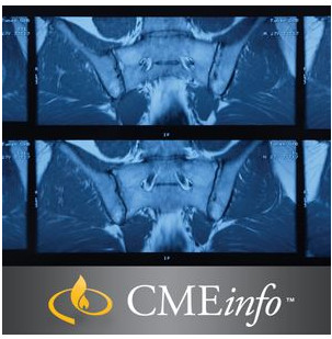
UCSF Musculoskeletal Imaging 2020
University of California San Francisco Clinical Update (SA-CME)
This exploration of musculoskeletal imaging delves into the anatomy and pathology of major joint conditions, cartilage lesions, bone tumors, and more.
Learn from the Experts
UCSF Musculoskeletal Imaging is a comprehensive state-of-the-art update on various aspects of musculoskeletal imaging. This well-planned course, directed by Ramya Srinivasan, MD, reviews pertinent anatomy and joint conditions for the shoulder, elbow, wrist, hip, ankle and foot, in addition to high-yield pathology topics such as musculoskeletal trauma, metabolic bone disease, arthritis and infection, and more. It will help you to better:
Topics/Speaker
Details : 23 Videos + 2 PDF
|
|
Related Products
CARDIOLOGY BOOKS
CARDIOLOGY BOOKS
Internal Medicine Videos
2022 AANEM Spring Virtual Conference Collection 2022 (CME VIDEOS)
Internal Medicine Videos
The International Congress Of Parkinson and Movement Disorder 2022 (MDS Congress) (CME VIDEOS)
GENERAL PEDIATRICS
Chestnet Pediatric Pulmonary Board Review On Demand 2022 (CME VIDEOS)
INTENSIVE CARE BOOKS
Chestnet Critical Care Board Review On Demand 2022 (CME VIDEOS)
Internal Medicine Books
Internal Medicine Books
The PassMachine Medical Oncology Board Review 2020 (v5.1) (Beattheboards) (Lectures)
Internal Medicine Books
8th Congress of the European Academy of Neurology – Europe 2022 (CME VIDEOS)
Internal Medicine Books
MD Anderson A Comprehensive Board Review in Hematology and Medical Oncology 2021 (CME VIDEOS)
Internal Medicine Videos
CARDIOLOGY BOOKS
Mayo Clinic Echocardiography Online Board Review 2022 (CME VIDEOS)
Internal Medicine Videos
Internal Medicine Books
Internal Medicine Books
The PassMachine Addiction Medicine Board Review 2022 (v3.1) (Beattheboards) (Lectures)
Internal Medicine Videos
Internal Medicine Videos
Internal Medicine Videos
Internal Medicine Videos
The Brigham Board Review and Comprehensive Update in Rheumatology 2022 (CME VIDEOS)
Internal Medicine Videos
Contemporary Issues in Breast Pathology uscap 2022 Items Included in the Purchase of this Course
Internal Medicine Videos
Internal Medicine Videos
Internal Medicine Videos
Internal Medicine Videos
Internal Medicine Videos
CHEST Advanced Critical Care Echocardiography Board Review Exam Course Virtual Event 2020
Internal Medicine Videos
Internal Medicine Videos
Internal Medicine Videos
Internal Medicine Videos
Cleveland Clinic Digestive Disease and Surgery Update OnDemand 2019
Internal Medicine Videos
GI BOARD REVIEW (The William M. Steinberg Board Review in Gastroenterology)
Internal Medicine Videos
Internal Medicine Videos
Johns Hopkins Review of Medical and Surgical Gastroenterology 2018 (Videos+PDFs)
Internal Medicine Videos
Internal Medicine Videos
Internal Medicine Videos
USCAP Tutorial in Pathology of the GI Tract, Pancreas and Liver 2019
Internal Medicine Videos
Internal Medicine Videos
The Brigham Board Review in Gastroenterology and Hepatology 2018
Internal Medicine Videos
Internal Medicine Videos
2019 Classic Lectures in Pathology What You Need to Know Pancreatobiliary Pathology
Internal Medicine Videos
2019 Classic Lectures in Pathology What You Need to Know Gastrointestinal Pathology
Internal Medicine Videos
Internal Medicine Videos
Internal Medicine Videos
Internal Medicine Videos
Internal Medicine Videos
AAFP FAMILY MEDICINE BOARD REVIEW SELF-STUDY PACKAGE – 13TH EDITION 2020
Internal Medicine Videos
Internal Medicine Videos
Internal Medicine Videos
Internal Medicine Videos
Internal Medicine Videos
Internal Medicine Videos
Internal Medicine Videos
Internal Medicine Videos
Internal Medicine Videos
Internal Medicine Videos
2019 Association for Community Health Improvement (ACHI) National Conference
Internal Medicine Videos
A Core Curriculum in Adult Primary Care Medicine 2018-2019 Lecture Series
Internal Medicine Videos
2019 Classic Lectures in Pathology What You Need to Know Endocrine Pathology
Internal Medicine Videos
Introduction to Adult EM Bootcamp + The Practice of Emergency Medicine (Hippo) 2020
Internal Medicine Videos
CCME The National Emergency Medicine Board Review course 2020
Internal Medicine Videos
Internal Medicine Videos
Internal Medicine Videos
Internal Medicine Videos
Internal Medicine Videos
ISICEM International Symposium on Intensive Care & Emergency Medicine 2020
Internal Medicine Videos
CCME Emergency Medicine & Acute Care: A Critical Appraisal Series 2020
Internal Medicine Videos
Internal Medicine Videos
Internal Medicine Videos
Internal Medicine Videos
CCME National Emergency Medicine Board Review Self-Study 2019 (Videos)
Internal Medicine Videos
Internal Medicine Videos
Internal Medicine Videos
The Passmachine Critical Care Medicine Board Review Course 2018
Internal Medicine Videos
Internal Medicine Videos
Internal Medicine Videos
Internal Medicine Videos
Internal Medicine Videos
Internal Medicine Videos
Internal Medicine Videos
Internal Medicine Videos
Internal Medicine Videos
Internal Medicine Videos
Internal Medicine Videos
The National Family Medicine Board Review Self-Study Course 2020
Internal Medicine Videos
The Brigham and Dana-Farber Board Review in Hematology and Oncology 2020 (Videos+PDFs)
Internal Medicine Videos
Internal Medicine Videos
Internal Medicine Videos
Internal Medicine Videos
The Brigham Board Review in Pulmonary Medicine 2020 (Videos+PDFs)
Internal Medicine Videos
Internal Medicine Videos
Internal Medicine Videos
Internal Medicine Videos
Internal Medicine Videos
Internal Medicine Videos
36th Annual UCLA Intensive Course in Geriatric Medicine and Board Review 2020 (Videos+PDFs)
Internal Medicine Videos
Need-to-Know Emergency Medicine: A Review for Physicians in a Hurry 2020 (Videos+PDFs)
Internal Medicine Videos
Internal Medicine Videos

