- 188 pages
- Publisher: Jaypee Brothers Medical Pub; 1 edition (August 30, 2011)
- Language: English
- Type : PDF . NVA , Epub
==========================+======================
Note : We will send ebook download link after confirmation of payment via paypal success
Payment methods: Visa or master card (Paypal)
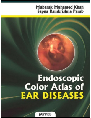
Endoscopic Color Atlas of Ear Diseases
$10
by Mubarak M. Khan (Author)
- Mubarak Muhamed Khan MBBS,DLODNB (ENT), Associate Professor, Department of Otolaryngology, MIMER Medical College, ConsultantOtolaryngologist, Sushrut ENT Hospital, Talegaon-D, Pune, Maharashtra, India
- Sapna Ramkrishna Parab MBBSMSDNB (ENT), Assistant Professor, Department of Otolaryngology, MIMER Medical College, Talegaon-D, Pune, Maharasthra, India
Endoscopic Color Atlas of Ear Diseases 1st Edition
By
Otology is an intriguing subdiscipline of otolaryngology—head and neck surgery. Ear disorders are one of the commonest diseases encountered in ear, nose and throat practice. The correct diagnosis of ear diseases requires a thorough knowledge of the normal anatomy and its alteration in pathological conditions. Middle ear anatomy is quite complex. Rendering a concrete picture of middle ear using only words had been always a challenging task. The constant effort to solve the doubts of the students in the best possible way was the driving force behind this atlas. With this backup in mind, this comprehensive atlas was developed with lucid and relevant text which addresses normal and diseased state of tympanic membrane, ear canal and middle ear. The atlas consists of a total of 271 colored photographs obtained by using 4 mm zero degree sinuscope. The clarity and the optics of the endoscope give greater information of the ear conditions contrary to the traditional outpatient Bull’s lamp or otoscopic examination. Before discussing the pathological conditions of the external and the middle ear, we have detailed the description of the normal tympanic membrane along with its variations. The appearance of the tympanic membrane is altered in various acute and chronic conditions affecting the middle ear. The alteration in its color, surface, intactness and position has been well illustrated in this atlas. The highlight of the atlas is unique approach of Exploration of the middle ear contents through subtotal perforation and its vast collection of pre and post operative images of Cartilage Tympanoplasty. This atlas will definitely supplement the standard ENT textbooks for a further in depth pictorial depiction of the ear disorders and thus facilitate proper diagnosis for dispensing appropriate treatment for otological disorders. We believe that this atlas will definitely be of immense help not only to the undergraduate and the postgraduate students, the ENT fraternity but also to general practitioners who also get a regular share of ENT patients. It is an update for the specialist treating otological pathologies as it is a “photogallery” of clinical findings accumulated over a period of time.
Product Details
Related Products
HEAD AND NECK SURGERY & OTOLARYNGOLOGY
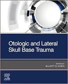
Otologic and Lateral Skull Base Trauma (True PDF from Publisher)
HEAD AND NECK SURGERY & OTOLARYNGOLOGY
HEAD AND NECK SURGERY & OTOLARYNGOLOGY
Neck Rejuvenation: Surgical and Nonsurgical Techniques (EPUB)
HEAD AND NECK SURGERY & OTOLARYNGOLOGY
HEAD AND NECK SURGERY & OTOLARYNGOLOGY
Jackson’s Local Flaps in Head and Neck Reconstruction (EPUB)
HEAD AND NECK SURGERY & OTOLARYNGOLOGY
HEAD AND NECK SURGERY & OTOLARYNGOLOGY
Atlas of Pediatric Head and Neck and Skull Base Surgery (EPUB)
HEAD AND NECK SURGERY & OTOLARYNGOLOGY
Contemporary Oral and Maxillofacial Surgery, 7th Edition (EPUB)
HEAD AND NECK SURGERY & OTOLARYNGOLOGY
HEAD AND NECK SURGERY & OTOLARYNGOLOGY
Oral and Maxillofacial Pathology, 5th Edition (True PDF from Publisher)
HEAD AND NECK SURGERY & OTOLARYNGOLOGY
HEAD AND NECK SURGERY & OTOLARYNGOLOGY
Textbook of Oral and Maxillofacial Surgery, 4th Edition (True PDF from Publisher)
HEAD AND NECK SURGERY & OTOLARYNGOLOGY
Technical Variations and Refinements in Head and Neck Surgery (Original PDF from Publisher)
HEAD AND NECK SURGERY & OTOLARYNGOLOGY
Chirurgie Maxillo-Faciale, 6th Edition (True PDF from Publisher)
HEAD AND NECK SURGERY & OTOLARYNGOLOGY
Atlas of Head and Neck Pathology, 4th Edition (True PDF from Publisher)
HEAD AND NECK SURGERY & OTOLARYNGOLOGY
Head and Neck Cancer Rehabilitation (True PDF from Publisher)
HEAD AND NECK SURGERY & OTOLARYNGOLOGY
HEAD AND NECK SURGERY & OTOLARYNGOLOGY
Head and Neck Cancer: Clinical Aspects (Original PDF from Publisher)
HEAD AND NECK SURGERY & OTOLARYNGOLOGY
HEAD AND NECK SURGERY & OTOLARYNGOLOGY
Multidisciplinary Head and Neck Reconstruction: A Defect-Oriented Approach (EPUB + Converted PDF)
HEAD AND NECK SURGERY & OTOLARYNGOLOGY
HEAD AND NECK SURGERY & OTOLARYNGOLOGY
HEAD AND NECK SURGERY & OTOLARYNGOLOGY
Thieme Dissector Volume 3: Head, Neck and Brain, 2nd Edition (Original PDF from Publisher)
HEAD AND NECK SURGERY & OTOLARYNGOLOGY
Pearls and Pitfalls in Head and Neck Pathology (Converted EPUB)
HEAD AND NECK SURGERY & OTOLARYNGOLOGY
2024 Practical Head & Neck Imaging: Mimics and Case-based Review – DocmedED (Videos)
HEAD AND NECK SURGERY & OTOLARYNGOLOGY
Operative Techniques in Spine Surgery, 3rd Edition (Original PDF from Publisher)
HEAD AND NECK SURGERY & OTOLARYNGOLOGY
2024 Multidisciplinary Head and Neck Cancers Symposium onDemand (Videos)
HEAD AND NECK SURGERY & OTOLARYNGOLOGY
Stell & Maran’s Head and Neck Surgery and Oncology, 6th edition (Original PDF from Publisher)
HEAD AND NECK SURGERY & OTOLARYNGOLOGY
HEAD AND NECK SURGERY & OTOLARYNGOLOGY
MAFAC Amsterdam 2023 – Cadaver Dissection Demonstrations & Lectures
HEAD AND NECK SURGERY & OTOLARYNGOLOGY
HEAD AND NECK SURGERY & OTOLARYNGOLOGY
Complex Head and Neck Microvascular Surgery: Comprehensive Management and Perioperative Care PDF
HEAD AND NECK SURGERY & OTOLARYNGOLOGY
Lateral Neck Swellings: Diagnostic and Therapeutic Challenges
HEAD AND NECK SURGERY & OTOLARYNGOLOGY
HEAD AND NECK SURGERY & OTOLARYNGOLOGY
Anatomy of Orofacial Structures: A Comprehensive Approach, 9th Edition
HEAD AND NECK SURGERY & OTOLARYNGOLOGY
HEAD AND NECK SURGERY & OTOLARYNGOLOGY
50 Landmark Papers every Thyroid and Parathyroid Surgeon Should Know
HEAD AND NECK SURGERY & OTOLARYNGOLOGY
Matrix Head and Neck Reconstruction: Scalable Reconstructive Approaches Organized by Defect Location
HEAD AND NECK SURGERY & OTOLARYNGOLOGY
HEAD AND NECK SURGERY & OTOLARYNGOLOGY
HEAD AND NECK SURGERY & OTOLARYNGOLOGY
Basic Guide to Oral and Maxillofacial Surgery (Basic Guide Dentistry Series)
HEAD AND NECK SURGERY & OTOLARYNGOLOGY
HEAD AND NECK SURGERY & OTOLARYNGOLOGY
HEAD AND NECK SURGERY & OTOLARYNGOLOGY
HEAD AND NECK SURGERY & OTOLARYNGOLOGY
HEAD AND NECK SURGERY & OTOLARYNGOLOGY
Otolaryngology: Current Concepts and Techniques in Head and Neck Surgery
HEAD AND NECK SURGERY & OTOLARYNGOLOGY
HEAD AND NECK SURGERY & OTOLARYNGOLOGY
Nanostructures for Oral Medicine (Micro and Nano Technologies)
HEAD AND NECK SURGERY & OTOLARYNGOLOGY
Kurzlehrbuch Hals-Nasen-Ohren-Heilkunde (UNI-MED Science), 4th Edition
HEAD AND NECK SURGERY & OTOLARYNGOLOGY
Postlaryngectomy voice rehabilitation with voice prostheses (UNI-MED Science)
HEAD AND NECK SURGERY & OTOLARYNGOLOGY
HEAD AND NECK SURGERY & OTOLARYNGOLOGY
HEAD AND NECK SURGERY & OTOLARYNGOLOGY
HEAD AND NECK SURGERY & OTOLARYNGOLOGY
HEAD AND NECK SURGERY & OTOLARYNGOLOGY
The Milan System for Reporting Salivary Gland Cytopathology, 2nd Edition
HEAD AND NECK SURGERY & OTOLARYNGOLOGY
HEAD AND NECK SURGERY & OTOLARYNGOLOGY
HEAD AND NECK SURGERY & OTOLARYNGOLOGY
Hair Cell Regeneration (Springer Handbook of Auditory Research, 75)
HEAD AND NECK SURGERY & OTOLARYNGOLOGY
HEAD AND NECK SURGERY & OTOLARYNGOLOGY
Soundscapes: Humans and Their Acoustic Environment (Springer Handbook of Auditory Research, 76)
HEAD AND NECK SURGERY & OTOLARYNGOLOGY
Simulation-Based Learning in Communication Sciences and Disorders: Moving from Theory to Practice
HEAD AND NECK SURGERY & OTOLARYNGOLOGY
Fundamentals of Anatomy and Physiology of Speech, Language, and Hearing
HEAD AND NECK SURGERY & OTOLARYNGOLOGY
HEAD AND NECK SURGERY & OTOLARYNGOLOGY
100 Complications of Otorhinolaryngology & Skull Base Surgery PDF & VIDEO
HEAD AND NECK SURGERY & OTOLARYNGOLOGY
The Art of Body Contouring: After Massive Weight Loss, 2nd edition Original PDF and Video
HEAD AND NECK SURGERY & OTOLARYNGOLOGY
HEAD AND NECK SURGERY & OTOLARYNGOLOGY
2023 Osteocom Hard and Soft Tissue Augmentation 2.0 – Luca De Stavola
HEAD AND NECK SURGERY & OTOLARYNGOLOGY
Sinus Grafting for Implant Reconstruction (Pikos Online MasterClass Series) – Michael A. Pikos
HEAD AND NECK SURGERY & OTOLARYNGOLOGY
Osteocom Full Tilted Implants Prosthetic and Surgical Protocols 2023
HEAD AND NECK SURGERY & OTOLARYNGOLOGY
HEAD AND NECK SURGERY & OTOLARYNGOLOGY
Optimizing Orthognathic Surgery: Diagnosis, Planning, Procedures (Epub & Converted to PDF)
HEAD AND NECK SURGERY & OTOLARYNGOLOGY
HEAD AND NECK SURGERY & OTOLARYNGOLOGY
2023 Classic Lectures in Head & Neck Imaging – A CME Teaching Activity
HEAD AND NECK SURGERY & OTOLARYNGOLOGY
Structure & Preservation Rhinoplasty Conference 2021 Istanbul
HEAD AND NECK SURGERY & OTOLARYNGOLOGY
HEAD AND NECK SURGERY & OTOLARYNGOLOGY
HEAD AND NECK SURGERY & OTOLARYNGOLOGY
HEAD AND NECK SURGERY & OTOLARYNGOLOGY
HEAD AND NECK SURGERY & OTOLARYNGOLOGY
Working with Voice Disorders, 3e (Original PDF from Publisher)
HEAD AND NECK SURGERY & OTOLARYNGOLOGY
Principles and Practices in Augmentative and Alternative Communication (Original PDF from Publisher)
HEAD AND NECK SURGERY & OTOLARYNGOLOGY
HEAD AND NECK SURGERY & OTOLARYNGOLOGY
HEAD AND NECK SURGERY & OTOLARYNGOLOGY
Jaypee’s Video Atlas of Operative Otorhinolaryngology and Head & Neck Surgery (Videos Only)
HEAD AND NECK SURGERY & OTOLARYNGOLOGY
Ear, Nose and Throat Simplified, 3rd edition (Original PDF from Publisher)
HEAD AND NECK SURGERY & OTOLARYNGOLOGY
ORL: Réussir son DFASM – Connaissances clés, 5th edition (Original PDF from Publisher)
HEAD AND NECK SURGERY & OTOLARYNGOLOGY
HEAD AND NECK SURGERY & OTOLARYNGOLOGY
Invasive Skull Base Mucormycosis New Perspectives (Original PDF from Publisher+Videos)
HEAD AND NECK SURGERY & OTOLARYNGOLOGY
Atlas of Facial Nerve Surgeries and Reanimation Procedures Original PDF
HEAD AND NECK SURGERY & OTOLARYNGOLOGY
Non-Surgical Rhinoplasty (The PRIME Series) (Original PDF from Publisher)
HEAD AND NECK SURGERY & OTOLARYNGOLOGY
Rinologia: Master Techniques In Otolaryngology – Head And Neck Surgery (Original PDF from Publisher)
HEAD AND NECK SURGERY & OTOLARYNGOLOGY
Secrets: Otorrinolaringologia, 4th Edition (Original PDF from Publisher)
HEAD AND NECK SURGERY & OTOLARYNGOLOGY
Otorrinolaringologia: Manual Prático em Cores (Original PDF from Publisher)
HEAD AND NECK SURGERY & OTOLARYNGOLOGY
HEAD AND NECK SURGERY & OTOLARYNGOLOGY
Current Opinion in Otolaryngology & Head and Neck Surgery 2022 Full Archives (True PDF)
HEAD AND NECK SURGERY & OTOLARYNGOLOGY
HEAD AND NECK SURGERY & OTOLARYNGOLOGY
Pediatric Otolaryngology: Practical Clinical Management,1st edition (EPUB)
HEAD AND NECK SURGERY & OTOLARYNGOLOGY
Color Atlas of Endo-Otoscopy: Examination-Diagnosis-Treatment, 1st edition (EPUB)
HEAD AND NECK SURGERY & OTOLARYNGOLOGY
HEAD AND NECK SURGERY & OTOLARYNGOLOGY
Manual of Eye, Ear, Nose, and Throat Emergencies (Original PDF from Publisher)

